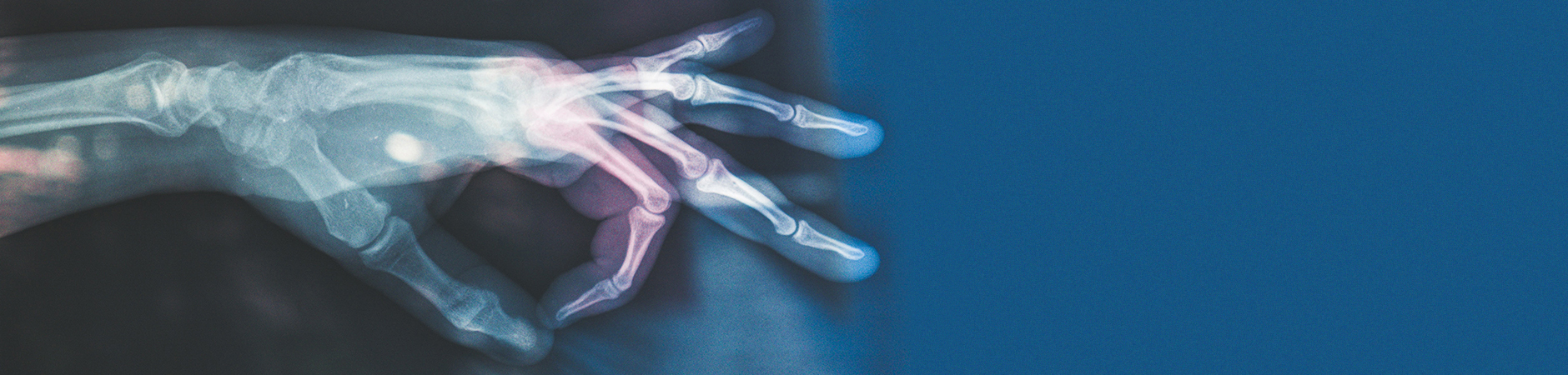
Medical physics (also called biomedical physics, medical biophysics or applied physics in medicine) is, generally speaking, the application of physics concepts, theories and methods to medicine or healthcare. Medical physics departments may be found in hospitals or universities. There are 4 main areas of medical physics speciality 1) radiation therapeutic physics, 2) medical imaging physics, 3) nuclear medicine physics and 4) health physics, which cover more than 90% of all medical physics activities.
Radiation therapeutic physicists work primarily in radiation oncology hospital departments, which specialize in cancer care. Radiation therapy (RT) is the most common treatment for cancer, being used in approximately 70% of all cancers either alone or combined with surgery or chemotherapy. It uses high-energy particles or waves, such as x-rays, gamma rays, electron beams, protons, carbon ions, to "kill" or "damage" cancer cells. There is a growing interest in the use of ion-beams (protons, carbon ions) for cancer therapy. The principal benefit of ion-beams are there finite range (or depth) in tissue, known as Bragg peak, where a significant amount of the radiation is deposited at the end of the track where the ions stop. The Bragg peak guarantees that healthy organs distal (deeper) to this peak receive very little or NO radiation, reducing significantly side effects. However, due to treatment and beam delivery uncertainties, it is not possible to accurately place the Bragg peak on the distal edge of the tumor. Thus, we voluntarily irradiate healthy surrounding organs to guarantee the tumor receives the needed radiation dose to sterilize the cancer. The Bragg peak 'uncertainty' reduces the clinical benefit of ion-beam radiotherapy.
Ongoing radiation oncology medical physics research in Heidelberg focuses on 1) developing novel imaging technologies to reduce the Bragg positioning uncertainty in patients using prompt gamma spectroscopy and Helium beam imaging for carbon ion therapy; 2) motion management technology for reducing effects during patient treatments, 3) incorporation of physical and mathematical models into the decision making process for radiotherapy, and 4) exploitation of the capabilities of the pixelized semiconductor detector Timepix for Helium and Carbon ion therapy. In addition, the molecular mechanism of DNA damage due to radiation is poorly understood. Thus, the field of radiation biology focuses on studying the biological effect on tissues of ionizing radiation.
Ion-beam therapy
Group J. Seco: In principle, ion-beam therapy offers a substantial clinical advantage over conventional photon therapy. This is because of the unique Bragg peak depth-dose characteristics, which can be exploited to achieve significant reductions in normal tissue doses proximal and distal to the target volume. These may, in turn, allow escalation of tumor doses and greater sparing of normal tissues, thus potentially improving local control and survival while at the same time reducing toxicity and improving quality of life. In the future, a more widespread use of ion-beam radiotherapy will make it possible to significantly improve cancer survival with minimal side effects. However, in order to take full advantage of ion-beam radiotherapy a better control is needed of the Bragg peak within the patient (cancer) and a better understanding of the radiation triggered DNA damage is required. Once we can control very accurately the positioning of the Bragg peak within the cancer to within 1mm, then it will be possible to reduce radiation side-effects, while simultaneously boosting the cancer with more radiation.
Our aims are to develop novel imaging technologies to reduce the Bragg peak positioning "uncertainties" for ion-beam radiotherapy, using Helium beam imaging and prompt gamma spectroscopy and to investigate the mechanism of radiation triggered DNA damage via reactive oxygen species (ROS). We are currently developing an high energy resolution detector system to perform spectroscopy of the prompt gamma radiation emitted during proton, Helium and Carbon ion therapy. We perform Monte Carlo radiation transport simulations to investigate the nuclear excited states produced by the different ion beams and to optimize the detection of the Prompt Gamma. Our aim is to perform online monitoring of the delivered Bragg peak during therapy.

Medical imaging physicists work primarily in radiology departments within an hospital, and specialize in early detection, tumor characterization, treatment guidance and response monitoring using morphological, functional, metabolic and molecular imaging. The enormous advances in the understanding of human anatomy, physiology and pathology in recent decades have led to ever-improving methods of disease prevention, diagnosis and treatment. Many of these achievements have been enabled, at least in part, by advances in 1) multi-slice computed tomography (CT), 2) magnetic resonance imaging (MRI), 3) positron emission tomography (PET) and 4) single photon emission tomography (SPECT). Major achievements in Heidelberg have centered on the development and evaluation of novel techniques in high- and ultrahigh-field MRI, dual-energy/spectral CT, contrast-enhanced ultrasound and dynamic PET.
Ongoing medical imaging research in Heidelberg focuses on evaluation of novel techniques in high- and ultrahigh-field MRI, dual energy/spectral CT, contrast-enhanced ultrasound and dynamic PET including hybrid imaging using new radiotracers. Ongoing research centers on 1) development of ultrahigh-field imaging devices, 2) photon-counting CT detectors, 3) simultaneous optical imaging and tomography with MRI, PET, SPECT and ultrasound, 4) chemical exchange saturation transfer (CEST) and NMR spectroscopy (MRS) with 1H and X nuclei, 5) diffusion weighted imaging (DWI) to gain insights into the structure of biological structures and 6) quantitative susceptibility mapping (QSM).
Group M. Ladd: In MRI, a significant difference encountered at ultra-high magnetic fields (= 7 Tesla) is that the RF wavelength inside the tissue can be shorter than the cross-sectional dimension of the human body, which implies that phase effects and wave propagation have to be accounted for. We are developing new RF technology based on multi-channel excitation coils to provide precise control over the RF field distribution. A 32-channel RF array for whole-body excitation at 7 Tesla has been developed, see illustration
Group L. Schad: We are working on several aspects of improved oncological radiotherapy treatment planning and monitoring by using physiological and functional imaging of CT, MRI and PET. One of our main research aspects lies in developing new MR techniques (23Na imaging, dynamic MRT, diffusion, perfusion, blood bolus tagging, BOLD MRI) for clinical use in therapy planning and monitoring. Another main aspect denotes imaging of hyperpolarized 3He in the human lung, as well as T2*- and T1-techniques for non-invasive measurement of tissue oxygenation and perfusion in the myocardium which is of general interest in radiology.


